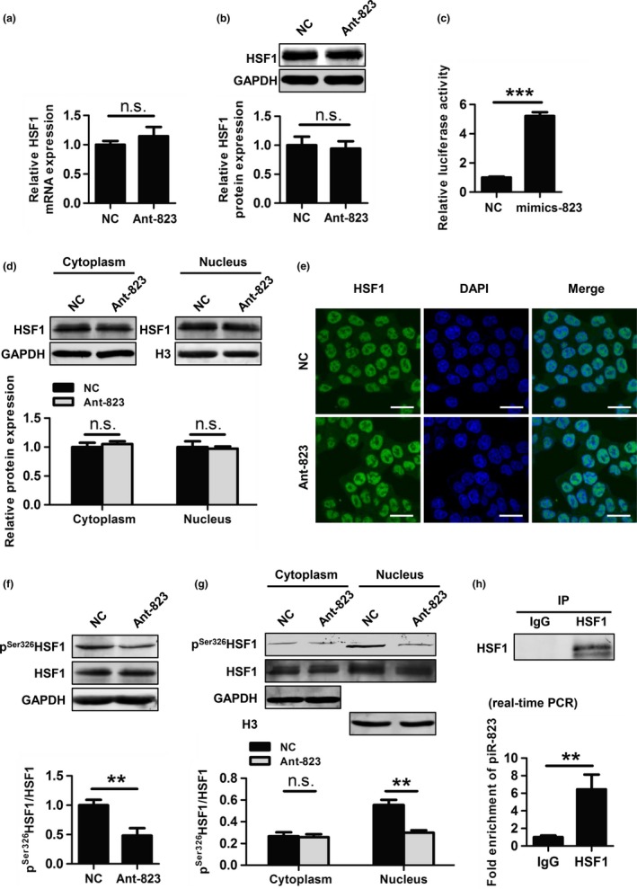
Figure 6.

piR‐823 regulates HSF1 activity by binding to HSF1 and modulating its phosphorylation at Ser326. (a, b) mRNA levels (a) and protein levels (b) of HSF1 was evaluated in HCT116 cells transfected with Ant‐823 or NC. (c) The transcriptional activity of HSF1 was assessed by dual‐luciferase reporter assay in HCT116 cells. (d, e) Nuclear translocation of HSF1 in HCT116 cells was evaluated by western blot analysis of cytoplasmic and nuclear extractions (d) and immunofluorescence staining for HSF1 (green) (e). Nuclear DNA was stained with DAPI (blue). Scale bar, 20 μm. (f) The expression of pS er326 HSF1 in HCT116 cells was assessed by western blot analysis. (d) Cellular localization of pS er326 HSF1 in HCT116 cells was determined by western blot analysis. (h) The interaction of piR‐823 with HSF1 in HCT116 cells was assessed by RIP assay using anti‐HSF1 or IgG antibody as a control. The eluted RNA was analyzed by real‐time PCR. Immunoprecipitation efficiency was assessed by western blot analysis. Data are shown as the mean ± SD from three independent experiments, **P < 0.01, ***P < 0.001, n.s. indicates no significance vs indicated groups.
