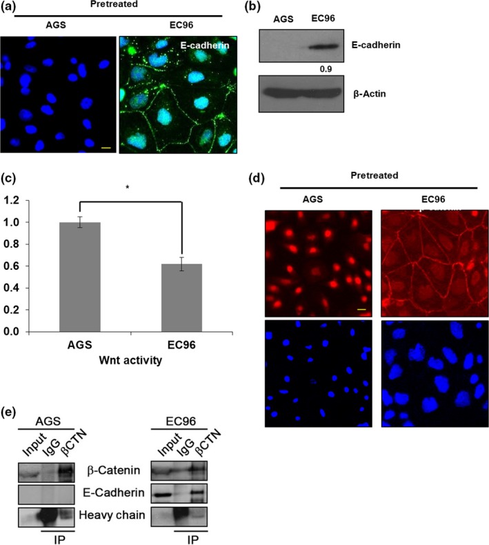
Figure 1.

E‐cadherin expression suppresses Wnt signaling in AGS gastric cancer cells. (a) AGS and EC96 cells were subjected to immunofluorescence staining for E‐cadherin. Scale bar = 30 μm. (b) Cell lysates were subjected to immunoblotting analysis of E‐cadherin and β‐actin. Band intensity was normalized to β‐actin. (c) Wnt signaling activities were measured by dual luciferase reporter assay. *P < 0.05. (d) Cells were subjected to immunofluorescence staining for β‐catenin. Scale bar = 30 μm. (e) Cell lysates were precipitated using an anti‐β‐catenin antibody, and the levels of E‐cadherin and antibody‐bound proteins were determined by immunoblot analysis using anti‐β‐catenin (βCTN) and anti‐E‐cadherin antibodies. IP, immunoprecipitant.
