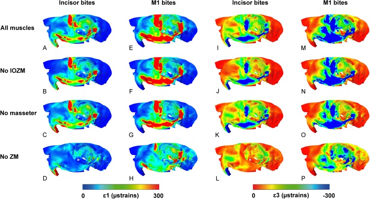
Figure 4. Predicted principal strains across the skull of Pedetes capensis during incisor and first molar biting.
(A–H) maximum (ε1) principal strains during incisor (A–D) and M1 (E–H) biting; (I–P) minimum (ε3) principal strains during incisor (I–L) and M1 (M–P) biting. (A, E, I, M) model with all masticatory muscles included; (B, F, J, N) model with IOZM excluded; (C, G, K, O) model with masseter excluded; (D, H, L, P) model with ZM excluded.

