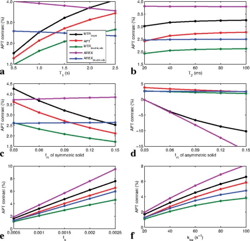
FIG. 3.
Simulated MTRasym, APT*, MTRdouble,vdc, AREX, and AREXdouble,vdc as a function of T1 (a), T2 (b), fm (c), fs (e), and ksw (f), respectively, with the symmetric-solid three-pool model (amide, symmetric semi-solid component, water), and as a function of fm (d) with the asymmetric-solid three-pool model (amide, asymmetric semi-solid component, water). Note that AREXdouble,vdc depends significantly only on the solute parameters fs and ksw, but not other tissue parameters, while all other APT imaging methods depend on several other tissue parameters such as T1, T2, and fm.

