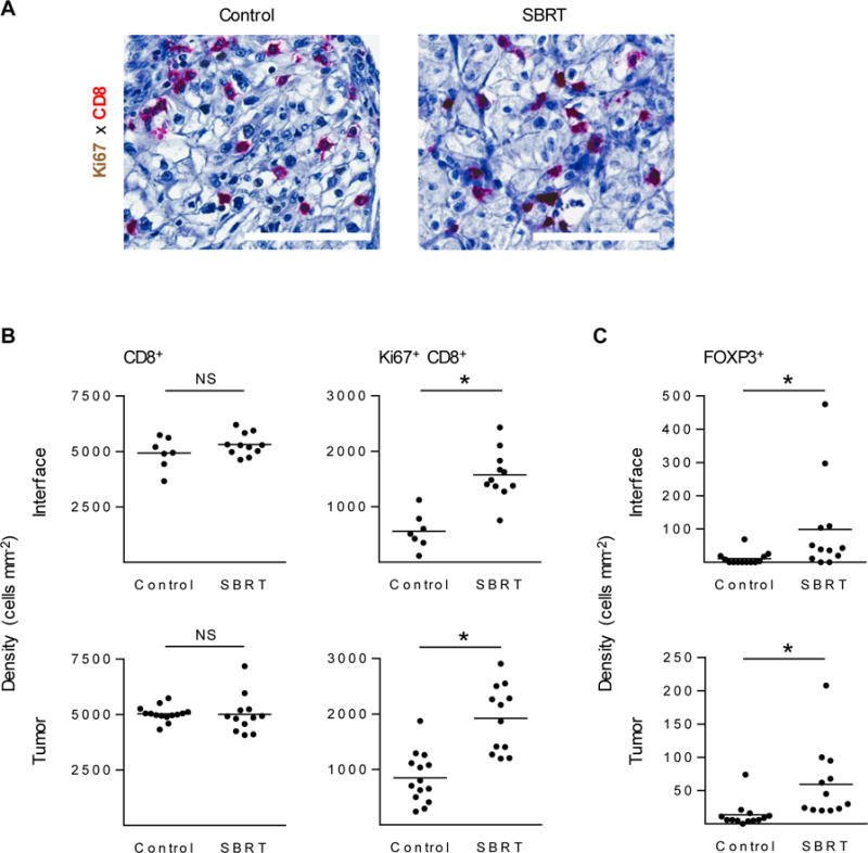
Figure 4. Tumors resected from SBRT-treated clear cell RCC patients have higher density of proliferating CD8+ T cells and FOXP3+ cells.

(A) IHC of CD8+ T cells (red) and Ki67+ (brown) in either control archival samples (left) or SBRT-treated (right) resected patient RCC tumors. Scale bars: 100 μm. Total CD8+ T cells numbers and Ki67+ CD8+ T cells were quantified by Aperio image analysis software (B). FOXP3+ cell density was evaluated in control and SBRT samples (C). n ≥ 7 patient samples per condition. *P < 0.05; NS, not significant; unpaired two-tailed Student’s t-test. Horizontal lines denote mean.
