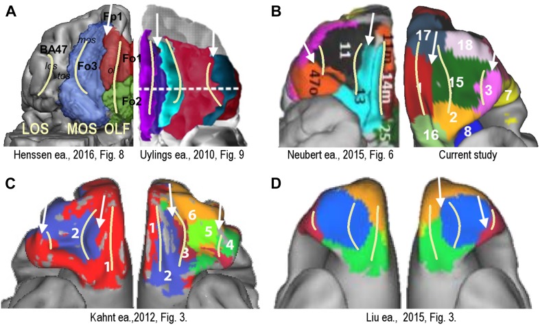
Fig. 5.
Orbital frontal surface comparison of parcellation schemes from different studies and different modalities with the group results from the current study. Yellow lines indicate location of major sulci: OLF olfactory sulcus, MOS medial orbital sulcus, LOS lateral orbital sulcus. White arrows indicate boundaries between delineated areas that possibly correspond to the significantly co-localized replicable boundaries found in this study (e.g., Fig. 2a). All pictures reproduced with permission. a Cytoarchitectonic parcellations: Henssen et al. (2016) (left) and Uylings et al. (2010) (right). b DWI probabilistic tractography: Neubert et al. (2015) (left); resting-state functional connectivity: data from the current study (right). c Group-wise resting-state functional connectivity from Kahnt et al. (2012): two cluster solution (left) and six cluster solution (right). d Local grey matter volume covariation: Liu et al. (2015), left and right hemisphere

