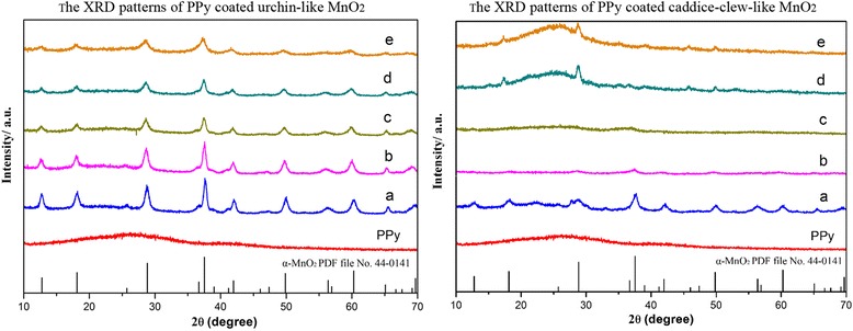
Fig. 5.

The XRD patterns of PPy-coated MnO2 samples. The left is (a) urchin-like MnO2 sample and (b) 10 μL, (c) 20 μL, (d) 30 μL, and (e) 50 μL PPy-coated. The right is (a) caddice-clew-like MnO2 sample and (b) 30 μL, (c) 50 μL, (d) 75 μL(e), and 100 μL PPy-coated
