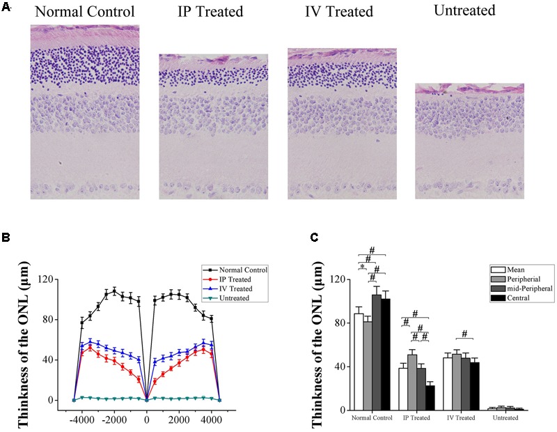
FIGURE 3.

Hydrogen rich saline induced effects on the retinal morphology of MNU administered rats. (A) The ONL architecture in the untreated group was terribly destroyed by MNU administration. Meanwhile, the photoreceptor in the IP and IV treated group were effectively rescued. (B) The ONL thickness of the IV treated group was ubiquitously larger than the IP treated group in the central, mid-peripheral, and peripheral regions. (C) The mean ONL thickness of the untreated group decreased significantly compared with the normal controls (P < 0.01); the mean ONL thickness of the IV treated group was significantly larger than the IP treated group (P < 0.01). The ONL thickness in the central region of the IP treated group was significantly smaller than the peripheral and mid-peripheral regions (P < 0.01); the ONL thickness of in the mid-peripheral region was significantly smaller than the peripheral region (P < 0.01). In IV treated group, the ONL thickness in the central region was significantly smaller than the peripheral region (P < 0.05) (All the values were presented as mean ± SD; ∗P < 0.05, #P < 0.01 for differences compared between the animal groups).
