Figure 7.
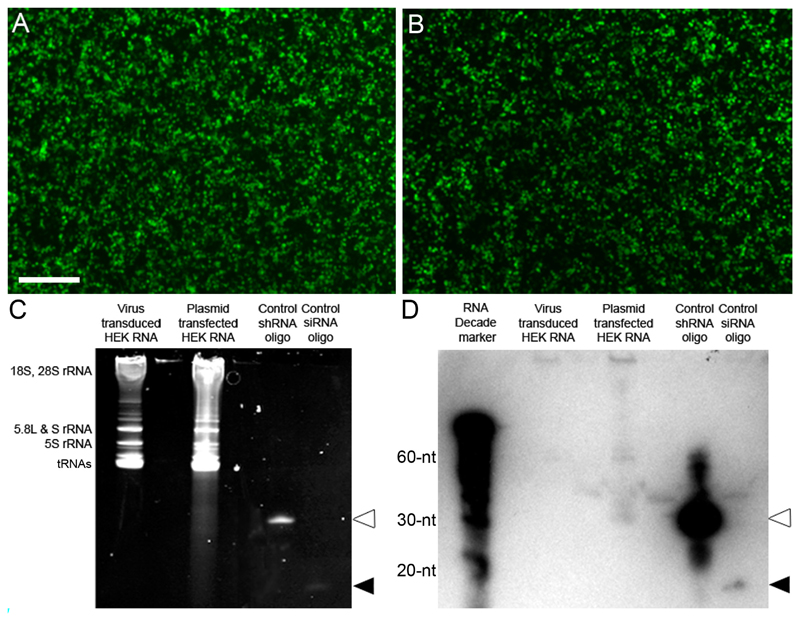
Northern blots do not detect a high level of shRNA expression. (A) Image of eGFP positive HEK cells that were efficiently transduced using Lenti-LINGO1-sh4 at an MOI 10. Scale bar: 500 μm. (B) Image of eGFP positive HEK cells that were efficiently transfected with the LINGO1-sh4 plasmid using PEI. (C) Image of the resolved RNA gel, the bands correspond to the different ribosomal RNA (rRNA) species or transfer RNAs (tRNAs) as labelled on the left side. The RNA resolved from the plasmid transfected HEK cells shows some smearing. The shRNA (white arrowhead) and siRNA (black arrowhead) positive controls can be seen, confirming the design and sensitivity of the probe (D) Image of the small transcript northern blot. Neither the shRNA nor siRNA was detected from the virus transduced HEK cell RNA. A faint band representing the shRNA was detected from the plasmid transfected HEK cell RNA but a band for the siRNA was not detected. Bands representing the shRNA (white arrowhead) and siRNA (black arrowhead) positive controls were detected.

