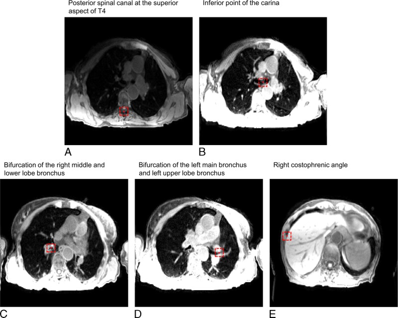
FIGURE 3.

An example of control point delineation for patient 8 on a snapshot image of 4D-T1w MRI. The control points (red circles) are contained within dashed red boxes to assist visualization. The presented windowing scheme was optimized by the radiation oncologist for viewing of the anatomical landmark and not for surrounding anatomy.
