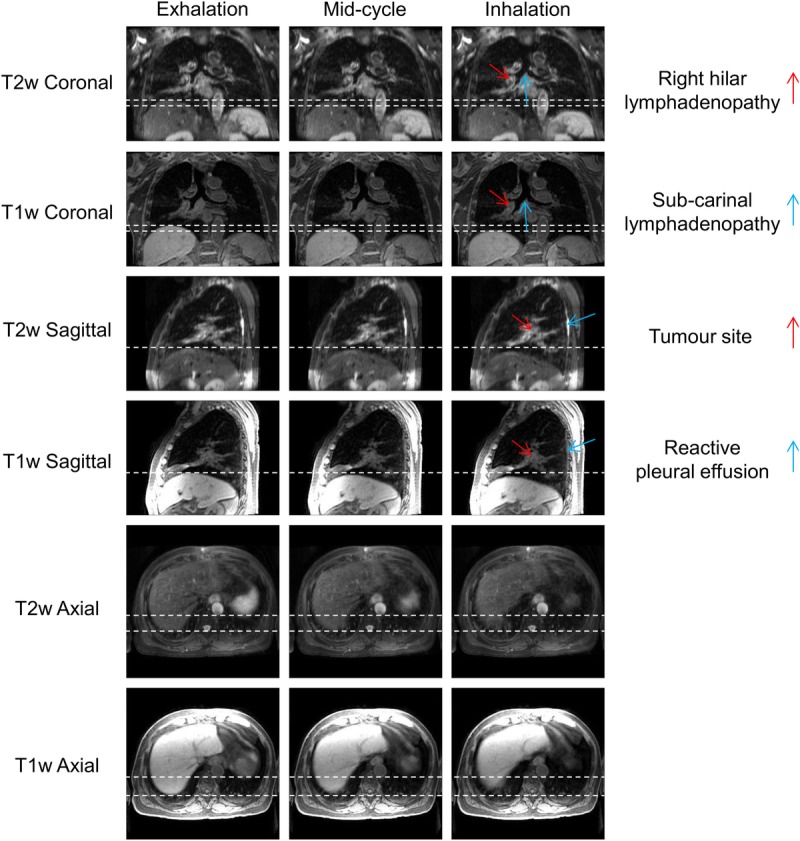
FIGURE 4.

Example T1w and T2w coronal, sagittal, and axial views at exhalation, midcycle, and inhalation respiratory phases for patient 7. This patient was diagnosed with T1aN2 primary lung squamous cell carcinoma and had a small benign reactive pleural effusion. The dashed white lines aid visualization of the diaphragm surface position. The respiratory pattern of T1w MRI is preserved in T2w MRI. The tumor site, lymphadenopathy, and reactive pleural effusion are shown more clearly in T2w MRI than T1w MRI.
