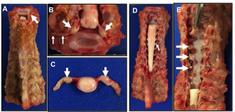
Figure 3.

Cervical to thoracic spine, dorsal view. A) The spine is sectioned from the head (at top of image) with the epaxial muscles and ribs removed. Note the small spinal nerve root (arrow) from the sectioning of the cervical spine. B) Cervical section through the spine (see top of A) that has been further dissected to reveal the dorsal root ganglia (large arrows) and spinal nerves (small arrows) extending from the spinal cord. C. Section of spinal cord and nerves with dorsal root ganglia (arrows). D) Dorsal view of cervical spine following laminectomy to reveal the spinal cord and nerve roots (arrow). E) The cervical spinal cord (see D) with spinal nerves was removed. Remnant spinal nerves can be seen exiting into the neuroforamina (arrows). Forceps (top of image) can be inserted into the neuroformina to grasp and remove the spinal nerve with dorsal root ganglia.
