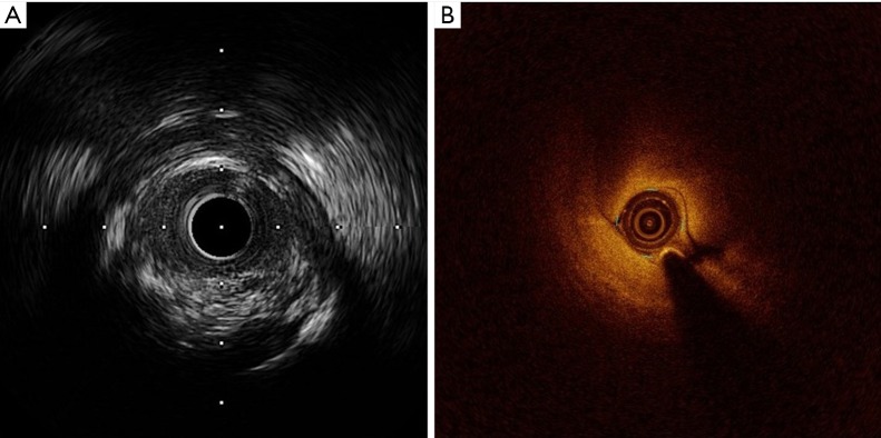
Figure 2.
IVUS (A) and OCT (B) images of proximal LAD stenosis. A fibrocalcific plaque causing severe stenosis was probed with both techniques. No evidence of intimal flap suggesting spontaneous coronary dissection was seen. IVUS, intravascular ultrasound; OCT, optical coherence tomography; LAD, left anterior descending artery.

