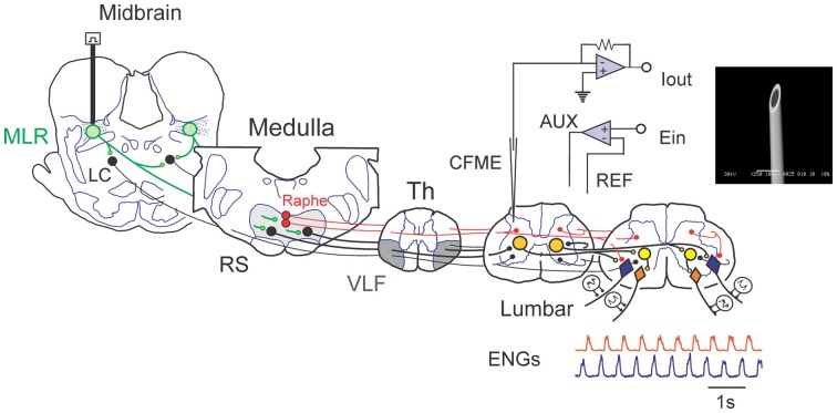
Figure 1.
Schematic view of experimental setup used to examine spinal monoamine release during mesencephalic locomotor region (MLR)-evoked fictive locomotion. Known neuronal projections from the MLR include the area of the noradrenergic locus ceruleus (LC), the serotonergic raphe nuclei in addition to the glutamatergic reticulospinal (RS) neurons within the medial reticular formation (Edwards, 1975; Steeves and Jordan, 1984; Sotnichenko, 1985). Axons of RS neurons descend via the ventrolateral funiculus (VLF) to innervate lumbar spinal locomotor interneurons comprising the central pattern generator (CPG; Steeves and Jordan, 1980; Noga et al., 2003). Fast cyclic voltammetry (FCV) scans within the lumbar spinal cord were applied throughout bouts of MLR evoked locomotion. All potentials (Ein) applied to the working carbon fiber microelectrode (CFME) are defined with respect to the reference (REF) electrode. If the potential applied to the CFME is different than the desired potential, then current is provided via the auxiliary (AUX) electrode (Ag/AgCl wire) to maintain the appropriate potential. Fictive locomotor activity is monitored by electroneurogram (ENG) recordings from hindlimb peripheral nerves. Inset: scanning electron micrograph of a CFME. The uninsulated carbon fiber (33 μm diameter) is visible at the beveled electrode tip.

