Figure 8.

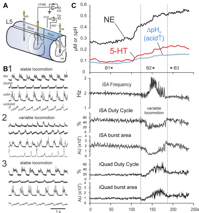
MLR-evoked spinal monoamine release is enhanced by monoamine metabolic and uptake inhibitors and shows modulation during locomotor transition periods. Release of NE and 5-HT in lamina VIII of the L5 segment (C) during dialysis of metabolic and uptake inhibitors pargyline (0.5 mM) and desipramine (0.1 mM) through dialysis probes placed 6 and 9 mm distant to the voltammetric electrode (experimental setup illustrated in (A)). Trial illustrated shows a 4 min portion, between 4.5 min and 8.5 min, of a 10 min locomotion trial. Locomotor activity for indicated periods in (C) are shown in (B,B1,2,3). At beginning of the illustrated sequence (as represented by locomotor period B1), extracellular levels of NE and 5-HT are already high (~275 and 100 nM, respectively) but are reasonably steady while locomotion parameters (frequency, duty cycle and burst area) remain stable (C). Transmitter levels dramatically change just prior to a period of variable locomotion (represented in B2) with changes in locomotor frequency and extensor/flexor burst area and burst duration (duty cycle). Following this period, monoamine levels regain a steady-state and locomotion is stabilized (B3). AU: arbitrary units. MLR stimulation parameters: 16 Hz, 160 μA, 1 ms duration pulses.
