
Figure 9.
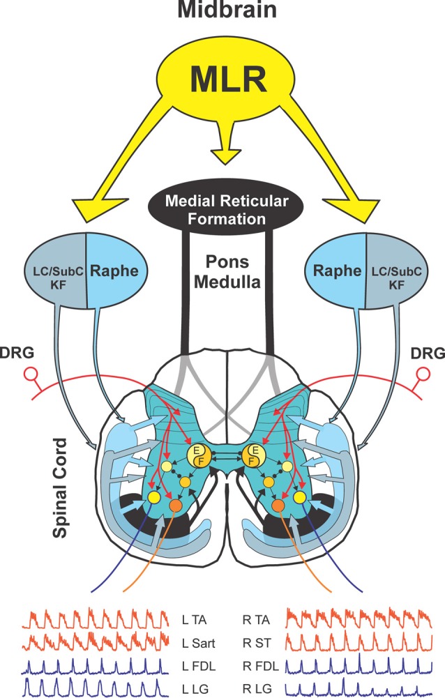
Model of descending locomotor pathways in the cat. Stimulation of the MLR activates reticulospinal neurons which project through the VLF to activate spinal locomotor CPG neurons, in part, by the release of excitatory amino acids. This pathway is considered to comprise the primary “command pathway” for the initiation of locomotion. MLR stimulation also activates in parallel, multiple monoaminergic descending pathways during centrally-generated fictive locomotion. This is evidenced by the rapid, widespread release of NE and 5-HT in the IZ/VH, in areas with centrally-activated locomotor neurons, and in the DH, where additional neurons are activated from afferent feedback from the moving limb during real locomotion. Extracellular monoamine levels are sufficient to activate monoaminergic receptors located on spinal locomotor-activated neurons indicating that extrasynaptic or volume transmission likely plays a significant role in the modulation/generation of locomotor activity. The flexor (F) and extensor (E) components of the locomotor CPG are activated/modulated by descending bilateral reticulospinal and monoaminergic projections as well as by crossed excitatory (▸) and inhibitory (•) segmental projections from the CPG opposite to it. Sensory afferents from skin and muscles innervate spinal neurons in DH, IZ and VH to fine-tune the locomotor step cycle. Details of the flexor and extensor components of the CPG are omitted in order to emphasize general interconnections between them and their target neurons. LC, locus ceruleus; SubC, subceruleus; KF, Kölliker-Fuse; DRG, dorsal root ganglia; L, left; R, right; TA, tibialis anterior, Sart, sartorius, FDL, flexor digitorum longus; LG, lateral gastrocnemius; ST, semitendinosus.