Figure 4.
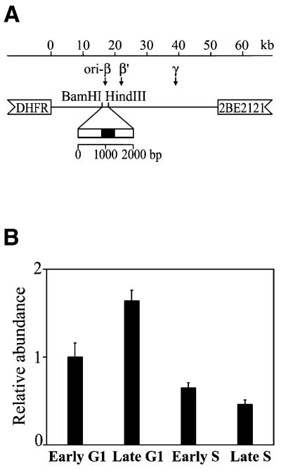
Dynamics of association of the ori-β to the nuclear matrix during the cell cycle of CHO cells. (A) Schematic representation of the position of the ori-β probe in the non-transcribed spacer 3′ of the DHFR gene in CHO cells. A 479 bp DNA fragment (filled box) was amplified by PCR and used as a probe. (B) Relative abundance of the DHFR ori-β in the matrix-attached DNA in CHO cells. Aliquots of matrix-attached DNA isolated from CHO cells synchronized at the indicated stages of the cell cycle were dot-blotted and hybridized with DNA probe in vitro labeled with [32P]dCTP. For determination of relative abundance the blots were rehybridized with genomic DNA. The autoradiographs were scanned and quantified and the ratios of probes to genomic DNA were calculated and presented in arbitrary units, assuming the early G1 ratio as 1.0. The results are means of five independent experiments and the standard deviations are shown with error bars.

