Figure 1.

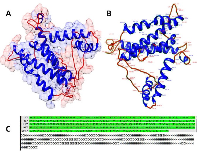
Visualizations of the predicted tertiary A: ribbon presentation with surface transparency (90%) & B: ribbon representation and secondary structure of the PLP protein (C) by Chimera software 1.8.

Visualizations of the predicted tertiary A: ribbon presentation with surface transparency (90%) & B: ribbon representation and secondary structure of the PLP protein (C) by Chimera software 1.8.