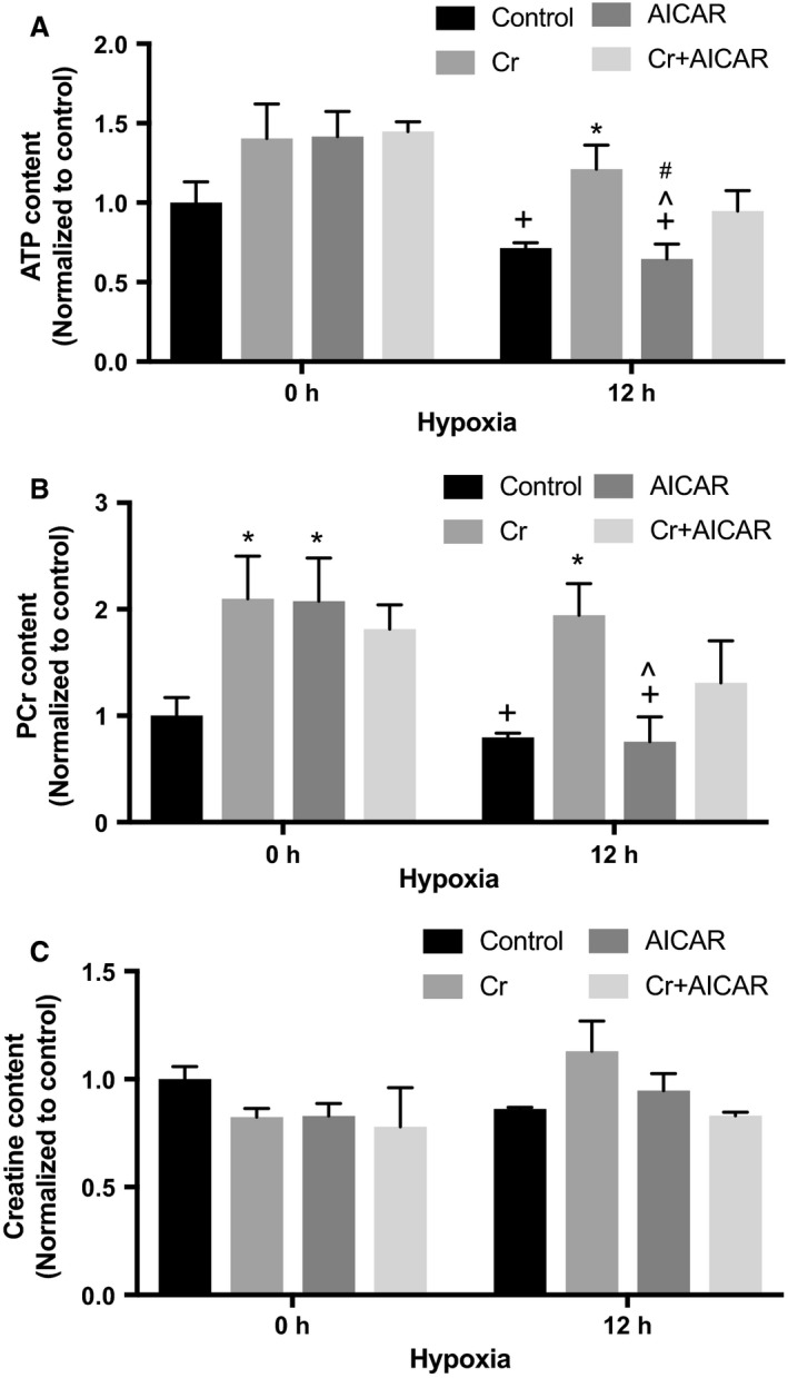
Figure 5.

Quantification of ATP, PCr, and Cr. RNCM cultures were pretreated with or without 0.5 mmol/L AICAR, 1 mmol/L Cr, or both for 24 h before exposure to 12 h of hypoxia. (A) ATP content was measured as described in Materials and Methods. Each bar represents the mean ± SEM of three independent measurements, normalized to control values. An “*” denotes a statistically significant difference compared with controls at the same hypoxia time point, a “+” denotes a statistically significant difference compared with Cr at 0 h, a “^” denotes a statistically significant difference compared with AICAR at 0 h, and a “#” denotes a statistically significant difference compared with Cr + AICAR at 0 h (two‐way ANOVA, P < 0.05, Tukey's test). (B) PCr content was measured as described in Materials and Methods. Each bar represents the mean ± SEM of three independent measurements, normalized to control. An “*” denotes a statistically significant difference compared with Control at the same hypoxia time point, a “+” denotes a statistically significant difference compared with Cr at 0 h, and a “^” denotes a statistically significant difference compared with AICAR at 0 h (two‐way ANOVA, P < 0.05, Tukey's test). (C) Cr content was measured as described in Materials and Methods. Each bar represents the mean ± SEM of three independent measurements, normalized to control (two‐way ANOVA, P = ns).
