
Fig. 1.
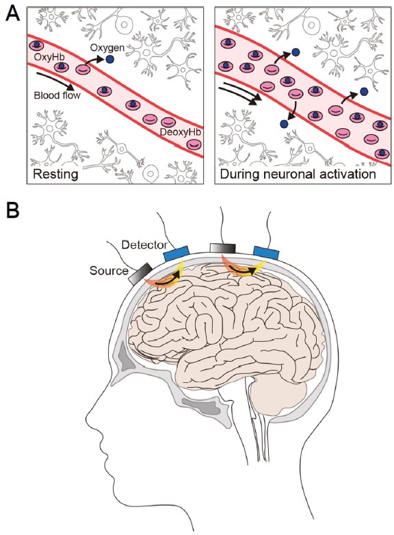
fNIRS imaging of cortical regions in humans.
(A) fNIRS uses near-infrared light to measures the relative changes in oxygenated (Oxy-Hb) and deoxygenated (Deoxy-Hb) hemoglobin in a brain region at baseline (“Resting”) and during neuronal activation. Oxy-Hb and deoxy-Hb are the main chromophores absorbing near-infrared light, and they exhibit distinct absorption spectra. (B) The surface of the head is irradiated with a combination of near-infrared wavelengths of light generated by the light source (“Source”). Photons returning to the surface of the head after traveling a banana-shaped path (in orange and yellow) in the tissues are captured by the photodetector (“Detector”) on the scalp. The number and array of the light source and photodetector on the head vary between studies.