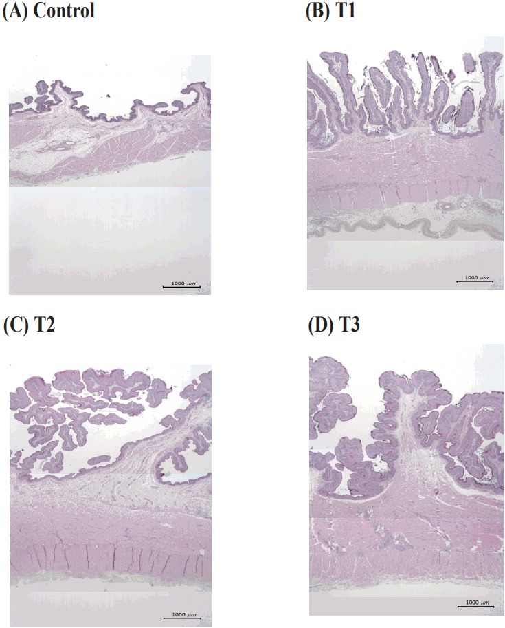
Figure 1.

Images from representative rumen papillae in six-month-old calves from different dietary treatment groups. (A) Control = maternal milk+Timothy hay, (B) T1 = milk replacer+concentrate, (C) T2 = milk replacer+concentrate+Timothy hay, (D) milk replacer+concentrate+30% starch. All sections of the ruminal papillae were stained with hematoxylin and eosin; ×12.5 magnification; bar = 1,000 μm.
