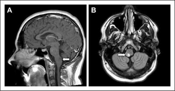
Figure 1.

Magnetic resonance images (MRIs) in an 11-year-old boy who has been followed closely, with no biopsy for confirmation and has not had any therapeutic intervention (Table 2). (A) T1-weighted sagittal non contrast image and (B) T1 axial flair image showing a dorsally exophytic medullary brainstem lesion.
