
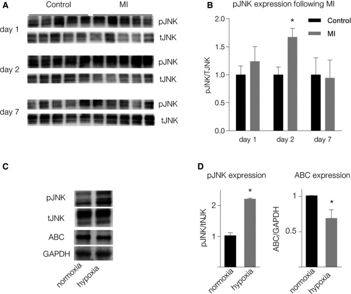
Figure 2. Non‐canonical WNT increases following MI.
-
ARepresentative Western blots of pJNK expression in the border zone of mouse hearts following MI.
-
BQuantification of pJNK expression (mean ± SD, N = 5, *P = 8.6E‐05, unpaired two‐sided Student's t‐test).
-
C, D(C) Representative Western blots and (D) quantification of pJNK (*P = 0.0001) and active β‐catenin (ABC) (*P = 0.001) expression in macrophages stimulated with supernatant of cardiomyocytes cultured under hypoxic conditions (unpaired two‐sided Student's t‐test, mean ± SD, N = 4).
Source data are available online for this figure.