Figure 2.

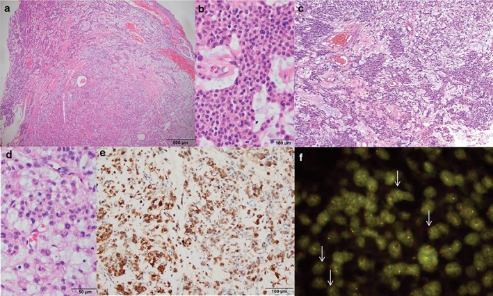
Pathological features. (a) A well‐demarcated cellular tumor in the myxoid stroma (hematoxylin end eosin [H&E] x 20). (b) Uniform tumor cells with a solid reticular growth pattern (H&E x 200). (c) Typical reticular features resembling extraskeletal myxoid chondrosarcoma (H&E x 100). (d) Mild nuclear atypia with multinucleated cells (H&E x 200). (e) Epithelial membrane antigen immunoreactivity (immunohistochemistry x 10). (f) Fluorescence in situ hybridization using the EWSR1 break‐apart probe revealed split red and green signals (arrows) suggestive of EWSR1 translocation.
