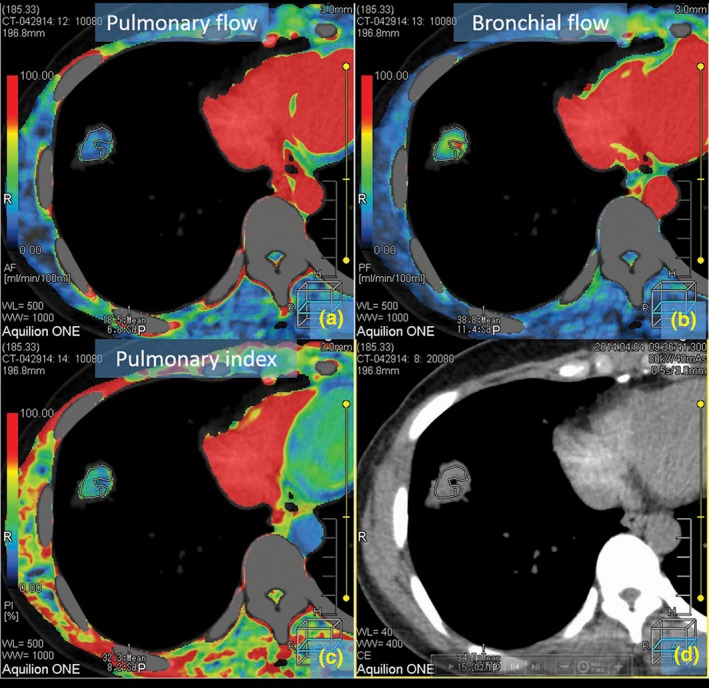
Figure 4.

Colored maps of dual‐input computed tomography perfusion in a 52‐year‐old man with peripheral adenocarcinoma in the right inferior lung. This nodule has a small cavity in the central portion. Air in the cavity may have resulted in miscalculation of the perfusion values. (a–c) show the maps color display of blood supply of the tumor for pulmonary flow (a), bronchial flow (b), and pulmonary flow (c). (d) shows computed tomography image without pseudo color processing.
