Fig. 3.
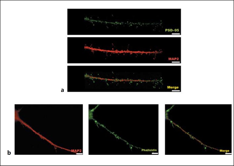
Visualization of dendritic filopodia and dendritic spines in low-density regions. Dendritic filopodia and dendritic spines can be directly analyzed in low-density regions by immunocytochemistry, without the need for transfection with a construct expressing a fluorescent reporter gene, which is required to highlight isolated neurons in high-density neuronal cultures. a Immunostaining with postsynaptic marker PSD-95 on a MAP2-positive dendrite. b Immunofluorescence-stained cells using phalloidin as F-actin stain to visualize dendritic filopodia and dendritic spines. Scale bars, 5 μm.

