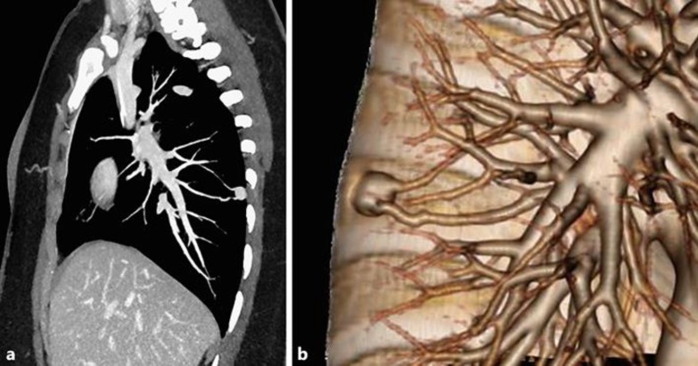
Fig. 2.
After 5 months of chemotherapy, the metastatic pulmonary nodules have decreased in size and are not enhancing, except for the nodule that developed arteriovenous fistula. A sagitally reformatted image with maximum intensity projection computed tomography (a) and a volume-rendered multidetector computed tomography image (b), showing communication of the nodule with the solitary feeding artery and a pulmonary draining vein, consistent with arteriovenous fistula.

