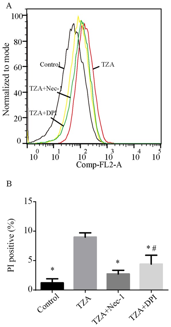
Figure 1.

PI+ HK-2 cells detected by flow cytometry in different groups. (A) TZA increased the percentage of PI+ HK-2 cells. Nec-1 and DPI exposure reduced the percentages of PI+ HK-2 cells induced by TZA. (B) The amount of the dead cells are presented as mean ± standard deviation. *P<0.05 vs. TZA group; #P<0.05 vs. control group. PI, propidium iodide; TZA, tumor necrosis factor-α + z-VAD-fmk + antimycin A; Nec-1, necrostatin-1; DPI, diphenyleneiodonium.
