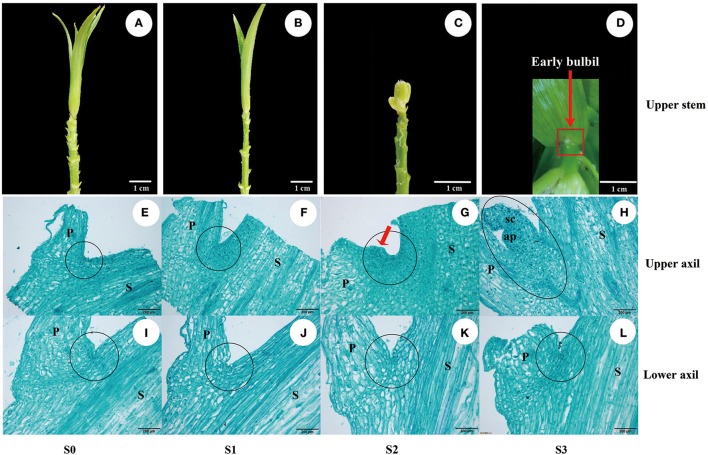
Figure 1.
Bulbil formation in L. lancifolium. (A–D) Stages of bulbil formation in the upper leaf axil: (A,B) S0 and S1 stages exhibiting apparent upper internodes; (C) S2 stage exhibiting swelling; (D) S3 stage exhibiting a white dot structure (early bulbil). (E–L) Microscopy images of paraffin-embedded samples of leaf axils located on the upper (E–H) and lower stem (I–L) from four stages (S0–S3). Scale bar for samples images = 1 cm; Scale bar for microscopy images = 200 μm. P, Petiole; S, stem; sc, scale; ap, apical point; black circle, leaf axil.

