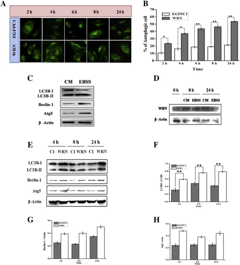
Fig. 3.

AG11395 cells were transfected with empty vector (EGFPC1) and vector containing full length WRN and starved with EBSS for 2 to 24 h. (A) Images of MDC staining taken with a fluorescence microscope under 40× magnification. (B) Graphical representation of % of autophagic cell during the time period (2 to 24 h). More than 500 cells per transfection were examined. * p < 0.05, ** p < 0.005, mean ± SD, n = 3. (C) WI-38 cells were transfected with full length WRN and allowed to express in complete medium (CM). After 24 h one set remains in complete medium (CM) and the other set was allowed for starvation for 24 h. (D) AG11395 cells were transfected with plasmid containing full length WRN and allowed to express WRN in complete medium (CM) for 24 h followed by starvation for 0, 8 and 24 h. Whole cell lysate was examined by immunoblotting with anti-WRN antibody. Here β-actin was used as loading control. (E) AG11395 cell was transfected with empty vector (EGFPC1) and full length WRN plasmid and starved after 24 h for 4, 8 and 24 h. Total cell lysate was immunoblotted with anti-LC3B, anti-beclin-1, and anti-Atg5 antibodies. Here β-actin was used as a loading control. (F–H) Band intensity was measured and graphically represents the ratio of LC3B-II and LC3B-I, beclin-1 and Atg5 at different time points respectively. ** p < 0.005, mean ± SD, n = 3.
