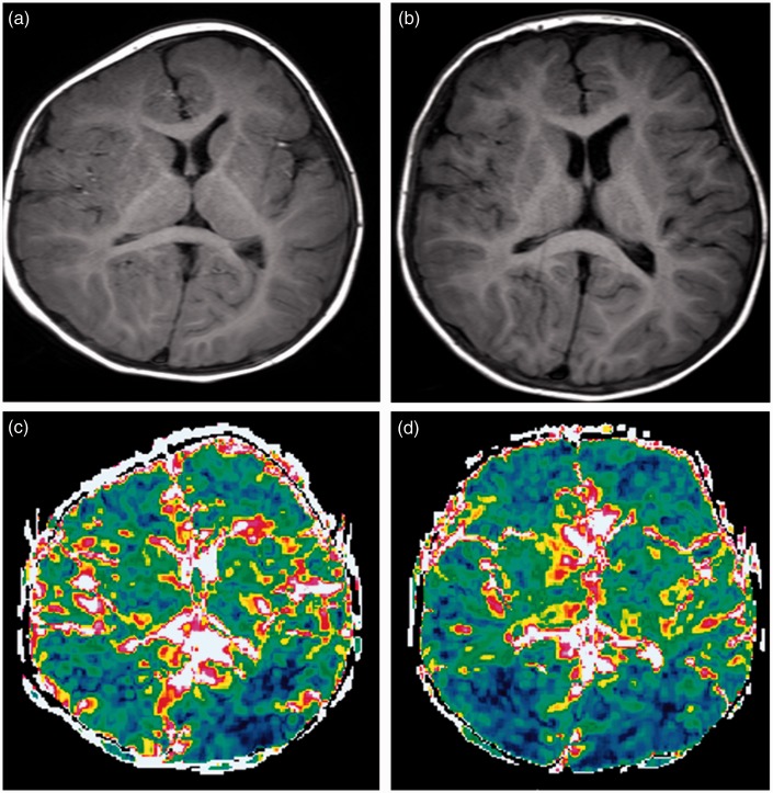
Fig. 3.
Patient 2. A girl with plagiocephaly (right coronal synostosis) and facial asymmetry. MRI was performed preoperatively at ten months (a) and postoperatively at one year five months (b). The right hemicalvarium was smaller than the left (a). After cranioplasaty, the calvarium is almost symmetrical (b). ΔADC maps obtained before (c) and after (d) the operation show changes of the ΔADC predominantly in the right frontal lobe. ΔADC values changed from 0.11 ± 0.020 to 0.15 ± 0.040 × 10⁻³ mm2/s.

