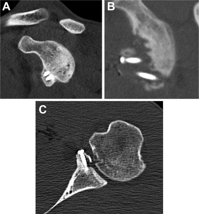
Figure 3.

(A) Sagittal 2-dimensional reconstruction of the left shoulder demonstrates the coracoid bone block in an optimal position, with nearly 100% bony bridging to the glenoid. Hardware is in place with minimal bone resorption. (B) Sagittal 2-dimensional reconstruction of the left shoulder of a different athlete that demonstrates significant bony resorption of the bone block. (C) Axial 2-dimensional image of the left shoulder that shows the bone block to be flush with the native glenoid with hardware in place but shows evidence of bony resorption lateral to the screw located within the bone block.
