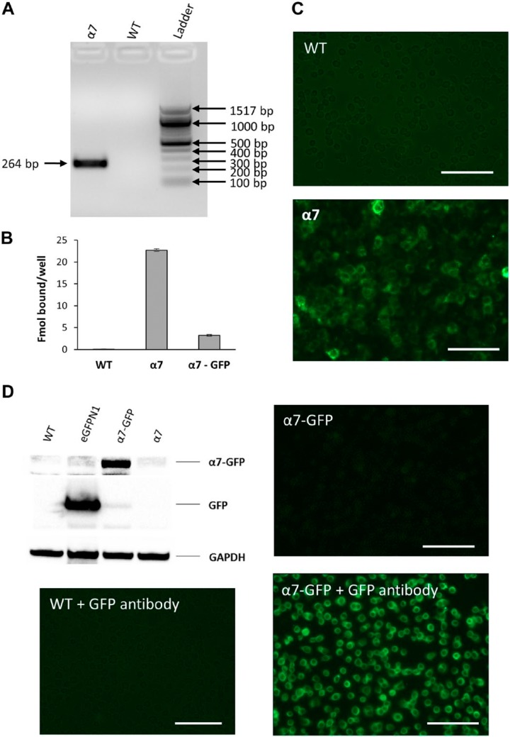
Figure 1.
Establishing rat α7 nAChR expression in GH4C1 cells. Expression of α7 nAChR RNA and protein levels in GH4C1 cells. (A) PCR analysis showing Chrna7 RNA expression in GH4C1-WT and α7 nAChR-transfected (α7) cells. α7 nAChR cDNA replicon band observed in the α7 cells at 264 bp, as expected, whereas no Chrna7 signal was observed in GH4C1-WT cells. (B) 125I-αBGT binding data comparing GH4C1-WT cells transfected with α7 or α7-GFP chimeras. α7- and α7-GFP-transfected GH4C1 cells showed significant binding whereas no binding was observed in untransfected cells. Bars in the figure represent the mean of specific binding and the error bars are the square root of the sum of the standard deviations squared for total and nonspecific binding. (C) Alexa Fluor 488-labeled αBGT binding showing fluorescence in α7-transfected GH4C1 cells compared with WT counterparts. (D) Western blot and immunofluorescence data to validate and compare WT with α7- and α7-GFP-transfected GH4C1 cells using a 1:2500 dilution of ab290 GFP antibody. A 25-kDa GFP band was observed only in the eGFPN1-transfected cells lysates (3-sec exposure). Similarly, an 80-kDa α7-GFP band was observed only in the α7-GFP transfected GH4C1 cell lysates (88-sec exposure). Forty μg of total protein was loaded in each lane and GAPDH was used as the loading control. WT GH4C1 cells do not show significant immunostaining with ab290 GFP antibody and Alexa Fluor conjugated rabbit secondary antibody. Immunofluorescence data showing only mild fluorescence in the α7-GFP-transfected cells that was increased significantly with GFP antibody staining. Scale bar, 40 μm. Abbreviations: nAChR, nicotinic acetylcholine receptors; WT, wild-type; GFP, green fluorescent protein.

