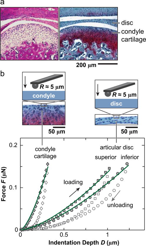
Fig. 1.

(a) Hematoxylin & Eosin (left) and Safranin-O/Fast Green (right) histology of sagittal sections showed the morphology of murine TMJ, highlighting the tissues of interest in this study: articular disc and mandibular condyle cartilage. (b) Representative indentation force versus depth, F-D, curves measured on the central regions of articular disc and mandibular condyle surfaces at 10 μm/s rate. The symbols are experimental data (density reduced for clarity), and solid lines are associated Hertz model fits to each of the loading F-D curve.
