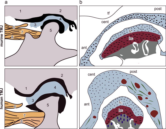
Fig. 7.

Schematics of the anatomical structure of murine versus human TMJs. (a) Anatomical structure of the TMJ: 1. articular eminence of temporal bone, 2. glenoid fossa of temporal bone, 3. anterior band of the articular disc, 4. posterior band of the articular disc, 5. mandibular condyle, 6. upper head of lateral pterygoid muscle, 7. upper head of the lateral pterygoid muscle connected to the mandible and 8. lower head of the lateral pterygoid muscle connected to the mandible condyle (adapted from (Suzuki and Iwata (2016)) with permission). (b) More detailed schematics of TMJ disc and condyle cartilage structure. In the murine TMJ, the articular disc has substantially smaller thickness compared to the condyle cartilage. This contrast is absent in larger animal TMJs (abbreviations, tf: temporal fossa, ant: anterior end, cent: central region, post: posterior end, fc: fibrocartilage, hc: hyaline cartilage). The tissue thickness values are: human: TMJ disc ~0.5–3 mm, condyle fibrocartilage layer: 400–600 μm, hyaline cartilage layer ~300–600 μm, as estimated in Bibb et al. (1993), Wright et al. (2016); murine TMJ disc ~30–60 μm, condyle fibrocartilage layer ~25–40 μm, hyaline cartilage layer ~90–120 μm.
