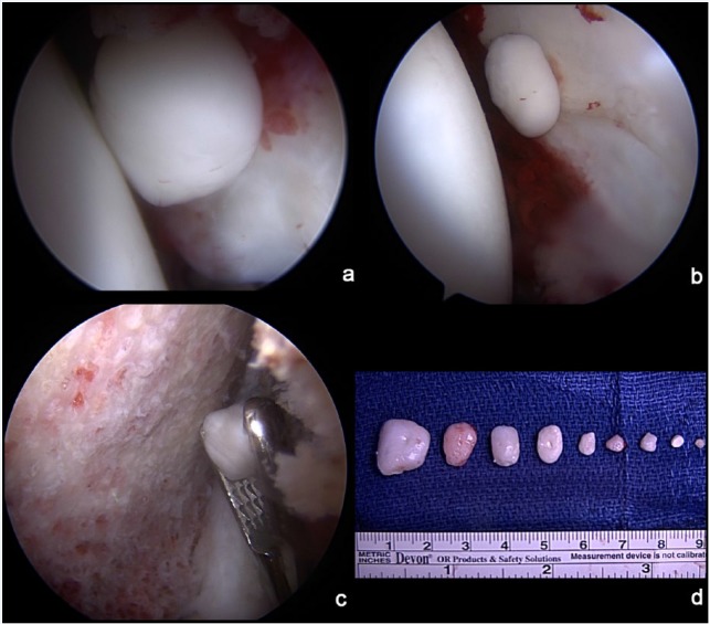
Figure 3.
Synovial chondromatosis. (a) A loose fragment is noted in the anterior aspect of the hip near the midanterior portal. (b) An additional fragment is noted laterally while viewing through the midanterior portal. (c) A large grasper is used to retrieve an additional loose body in the peripheral compartment along the femoral neck. (d) Gross view of the multiple loose fragments that were removed.

