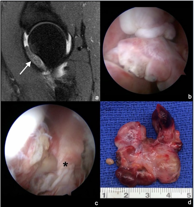
Figure 4.
Pigmented villonodular synovitis (PVNS). (a) Sagittal T2-weighted magnetic resonance image demonstrating the intra-articular soft tissue mass along the anterior femoral neck (arrow). (b) Visualization of the lobular, nodular PVNS mass. (c) A stalk is noted that connects the mass with the hip synovium of the anterior capsule (*). (d) Gross specimen demonstrating the multinodular and lobular nature of the PVNS mass.

