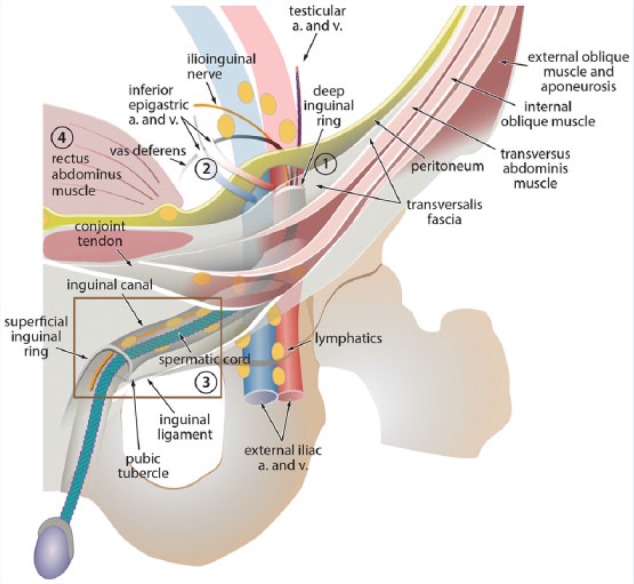
Figure 1.

Diagram of the inguinal canal (IC) and its contents. The deep inguinal ring is formed by the transversalis fascia, and the IC is lined by the same layers that line the abdominal wall. The external superficial ring is a triangular opening in the oblique aponeurosis. The inferior epigastric artery (a.) and vein (v.) originate from the external iliac artery and vein and lie medial to the internal inguinal ring. Locations of the abdominal wall hernias in relation to the IC are as follows: Indirect inguinal hernias lie lateral to the inferior epigastric arteries (1); direct inguinal hernias lie medial and inferior to the inferior epigastric vessels (2); femoral hernias lie inferior and medial to the femoral vessels (3); and spigelian hernias lie lateral to the rectus abdominus muscle (4). Reproduced with permission from Revzin MV, Ersahin D, Israel GM, et al. US of the inguinal canal: comprehensive review of pathologic processes with CT and MR imaging correlation. Radiographics. 2016;36:2028-2048.
