Figure 5.
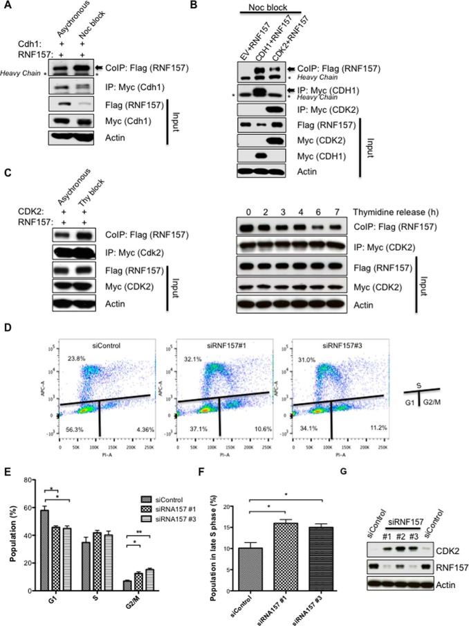
RNF157 role within the cell cycle. A, FLAG-RNF157 was co-transfected with Myc-CDH1 in HeLa cells and co-immunoprecipitated with Myc-CDH1 after DMSO or nocodazole (Noc) treatment. Lysates were subjected to immunoblotting using FLAG, Myc, and actin antibodies. B, Western blotting of RNF157 co-immunoprecipitated with CDH1 or CDK2 from HeLa cells arrested in G2/M by nocodazole. C, FLAG-RNF157 co-immunoprecipitated with Myc-CDK2 from HeLa cells with/without double thymidine (Thy) block. Western blot analysis was performed as in A. Cells were transfected and arrested with thymidine as in the left panel and then released into fresh medium for the times indicated. Western blots of FLAG-RNF157 co-immunoprecipitated with Myc-CDK2 were analyzed with the antibodies as indicated. D, analysis of cell cycle progression of HeLa cells transfected with control siRNA or two different siRNAs against RNF157. Samples were stained with the Click-iT EdU Assay kit using the manufacturer's recommended protocols 4 days post-transfection, and their DNA content was determined by flow cytometry. The plots show cells in different cell cycle phases. Cell cycle analysis was performed by gating G1, S, and G2/M on propidium iodide (PI-A) for DNA content and on Alexa Fluor 647 (APC-A) for EdU incorporation. E and F, quantification of the cell cycle analysis represented in D. Data represent means ± S.D. (error bars) obtained from three independent experiments to indicate a statistically significant result. p values are designated with asterisks as follows: *, p ≤ 0.05; **, p ≤ 0.01. G, HeLa cells were transfected with control siRNA or three different siRNAs against RNF157 for 4 days. The lysates were then subjected to immunoblotting to detect the endogenous proteins of interest by using the antibodies indicated. IP, immunoprecipitation, CoIP, co-immunoprecipitation.

