Figure 1.
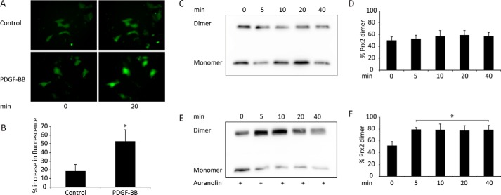
Prx2 dimer formation is induced by PDGF-BB treatment in MEF cells pretreated with auranofin. A, representative images of MEF cells expressing cytosolic HyPer after 0 and 20 min without treatment (Control) or treatment with 100 ng/ml PDGF-BB. B, change in fluorescence over 20 min in untreated or PDGF-BB–stimulated Hyper-expressing MEF cells. Data from 19 transfected cells were analyzed in five different experiments (mean ± S.E.; *, p < 0.05). C, Prx2 immunoblot of non-reducing SDS-PAGE–resolved lysates from MEF cells treated with 50 ng/ml PDGF-BB for the indicated times. D, quantification of Prx2 dimers (percentage of total Prx2) from densitometry analyses of blots represented in C (n = 7). E, immunoblot as in C of lysates from cells pretreated for 1 h with the TrxR1 inhibitor auranofin (1 μm) before PDGF-BB treatment. F, quantitation of Prx2 dimers in auranofin-treated cells determined as in D (mean ± S.E.; *, p < 0.05).

