Figure 4.
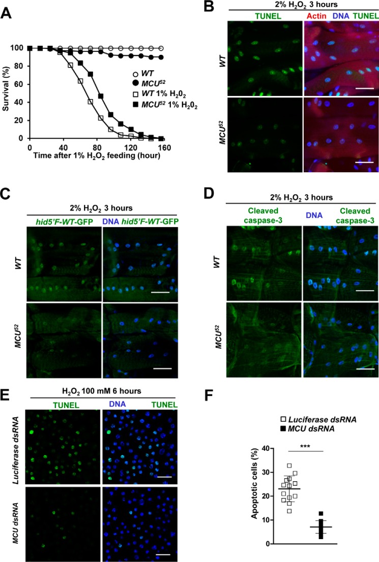
MCU mediates oxidative stress-induced apoptosis. A, survival curves of wild-type and MCU52 male flies on 1% H2O2-containing food (n = 133–149). Log-rank test: p < 0.0001. B–D, detecting apoptotic cells in larval muscle of wild-type and MCU52 flies after treatment of 2% H2O2 for 3 h. The assays were repeated >3 times. B, left panels, TUNEL signals; right panels, merged images of TUNEL (red), Hoechst (blue), and phalloidin (green) staining. C, left panels, GFP reporter expression from hid5′F-WT enhancer; right panels, merged images of GFP reporter (green) and Hoechst (blue) staining. Genotypes are WT (hid5′F-WT-GFP/+) and MCU52 (hid5′F-WT-GFP/+;MCU52/MCU52). D, left panels, cleaved caspase-3; right panels, merged images of cleaved caspase-3 (green) and Hoechst (blue) staining. E, TUNEL assays after treatment of 100 mm H2O2 for 6 h in S2 cells transfected with luciferase dsRNA or Drosophila MCU dsRNA. The TUNEL assay was repeated >3 times. F, the proportion of apoptotic nuclei in S2 cells transfected with luciferase dsRNA or Drosophila MCU dsRNA (n = 12–13). Scale bars, 50 μm (B–D) or 20 μm (E). ***, p < 0.001. Error bars, S.D.

