Fig. 2.
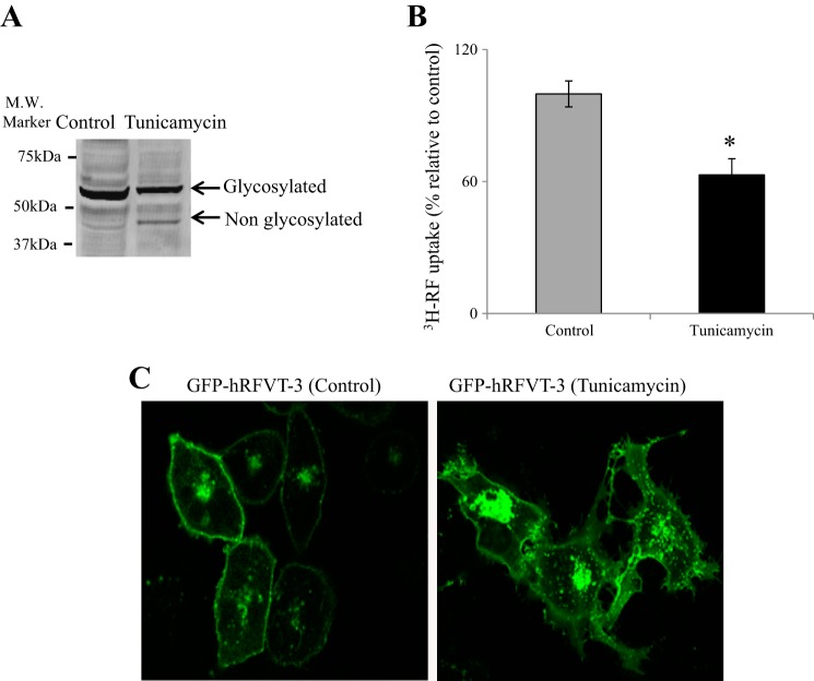
Effect of tunicamycin treatment on hRFVT-3 properties in HuTu-80 cells. A: Western blot analysis after tunicamycin treatment of green fluorescent protein (GFP)-hRFVT-3-expressing HuTu-80 cells. Western blotting was performed as described in materials and methods. The images are representative of three independent experiments with similar results. B: effect of tunicamycin treatment on riboflavin (RF) uptake. Uptake of [3H]RF (14 nM) was performed in Krebs-Ringer (K-R) buffer (pH 7.4) at 37°C for 3 min on GFP-hRFVT-3-expressing HuTu-80 cells with or without tunicamycin treatment (2 µg/ml, 24 h). Data are means ± SE of multiple determinations from at least four independent experiments. *P < 0.01. C: lateral (xy) images of GFP-hRFVT-3-expressing HuTu-80 cells: untreated control and treated with tunicamycin (2 µg/ml, 24 h).

