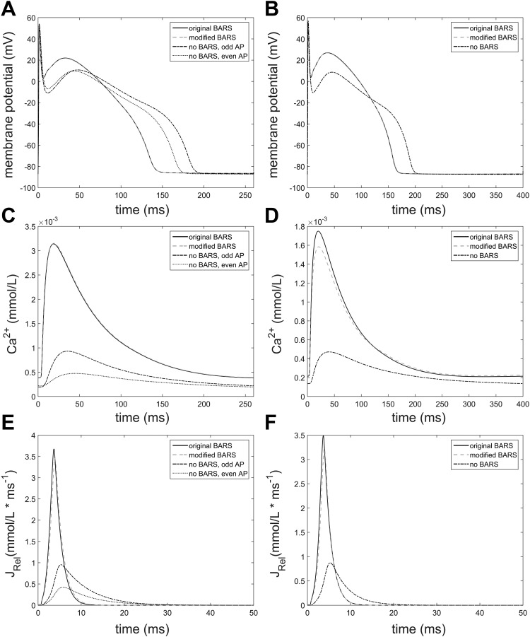
Fig. A2.
Comparison of the original and modified model of β-AR stimulation-dependent RyR2 regulation in a NZ cell, also showing traces from a NZ cell without β-AR stimulation. A, C, and E contain traces of membrane potential, free intracellular Ca2+, and Ca2+ release flux from the SR (having different x-axis) in a cell paced at a BCL of 260 ms. As the cell without β-AR stimulation manifests alternans, both alternating beats are shown. B, D, and F show the same variables but at a BCL of 400 ms, when no alternans is present.

