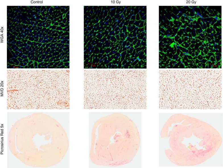
Fig. 7.
Myocardial histopathology. Top: representative examples of cardiomyocyte size [wheat germ agglutinin (WGA)-stained LV sections at ×40 magnification, short-axis myocyte orientation) for control, 10-Gy, and 20-Gy groups. Middle: representative examples of MVD (CD34-stained LV sections, ×20 magnification) for control, 10-Gy, and 20-Gy groups. Bottom: representative examples of myocardial fibrosis (picrosirius red-stained heart sections, ×5 magnification) for control, 10-Gy, and 20-Gy groups.

