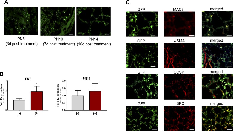
Fig. 2.
Viral expression in lung tissues. Pups were exposed as described, treated with a single dose of adeno-associated 9 (AAV9)-miR29b(+) or control(−) at postnatal day (PN) 3, euthanized at PN6, PN10, or PN14, and tissues were fixed (PN6 and PN10 lungs were not inflated because of pup size). A: sections were probed with anti-GFP antibody and immunofluorescence was visualized. Bars = 100 μm; photos were taken at ×200. B: additional lung tissues were harvested and analyzed by quantitative PCR analysis of miR-29b-3p at PN7 or PN14. Data were analyzed by t-test; n = 4 per group; *P < 0.05 different than control(−). C: coimmunofluorescence was assessed in tissues obtained at PN14. Bars = 100 μm in all images; photos were taken at ×400.

