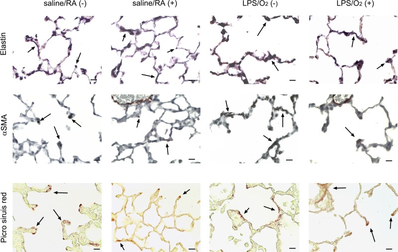
Fig. 5.
Immunohistochemical assessment of matrix proteins. Lung tissue from mice exposed to saline/RA or LPS/O2 and treated with a single dose of AAV9-miR-29b(+) or control virus(−) at PN3 were isolated and formalin fixed at PN28. Sections were stained with Hart’s stain for elastin (A), immunohistochemistry for α-smooth muscle actin (α-SMA; B), and Picrosirius red for collagen (C). Photomicrographs were taken at ×400; bars = 50 μm.

