Fig. 3.
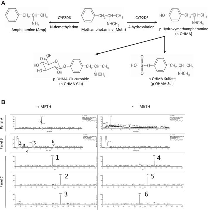
METH is metabolized by pulmonary microvascular endothelial cells (PMVECs) into p-hydroxymethamphetamine (p-OHMA). A: diagram illustrating the phase I metabolism of METH into amphetamine (Amp) and p-OHMA. p-OHMA could be further metabolized into p-OHMA glucuronide (p-OHMA-Glu) and p-OHMA sulfate (p-OHMA-Sul) in the liver to increase solubility and facilitate urinary excretion. B: liquid chromatography (LC)-mass spectrometry (MS) study of PMVECs treated with METH for 4 h. Analysis of cell lysates demonstrated at least 5 OHMA metabolites (peaks 2–6). Panel A represents full-scan LC-MS chromatograms of treated and untreated samples. Panel B represents collision-induced dissociation chromatograms (MS2 m/z 166.2 @CID 35.00) of hydroxylated methamphetamine metabolites in the same samples. Panel C represents MS-MS spectra of METH hydroxymetabolites (peaks 2–6). Peak 1 is a nonspecific endogenous component also found in unstimulated PMVECs.

