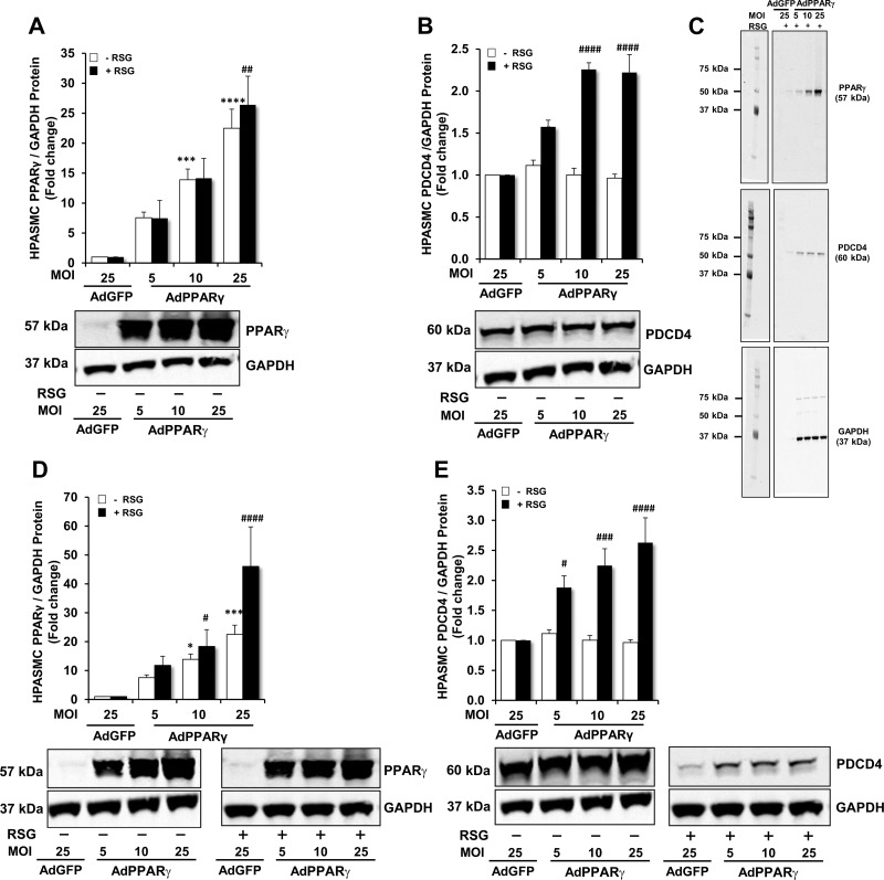
Fig. 4.
Time- and dose-dependent effects of PPARγ ± rosiglitazone (RSG) on PDCD4 and PPARγ protein expression. HPASMC monolayers were propagated in a cell culture incubator at 37◦ C until reaching 60–80% confluence. Selected HPASMCs were transfected with AdGFP [25 multiplicity of infection (MOI)] or escalating doses of AdPPARγ (5–25 MOI) for 6 h. After 24 h, RSG (10 μM) or an equal volume of DMSO was added to the cell culture media. Following the conclusion of 48- or 72-h incubation periods, total protein was isolated from monolayer lysates. A and B: PPARγ or PDCD4 protein levels were detected in HPASMCs transfected with AdGFP or AdPPARγ ± RSG for 48 h. C: full-length representative immunoblots are shown to demonstrate PPARγ, PDCD4 and GAPDH proteins in HPASMCs transfected with AdGFP or AdPPARγ + RSG for 48 h. D and E: PPARγ or PDCD4 protein levels were detected in HPASMCs transfected with AdGFP or AdPPARγ ± RSG for 72 h. Representative immunoblots are displayed in A, B, D, and E; n = 6. *P < 0.05 vs. AdGFP (−RSG); ***P < 0.001 vs. AdGFP (−RSG); ****P < 0.0001 vs. AdGFP (−RSG); #P < 0.05 vs. AdGFP (+RSG); ##P < 0.01 vs. AdGFP (+RSG); ###P < 0.001 vs. AdGFP (+RSG); ####P < 0.0001 vs. AdGFP (+RSG).

