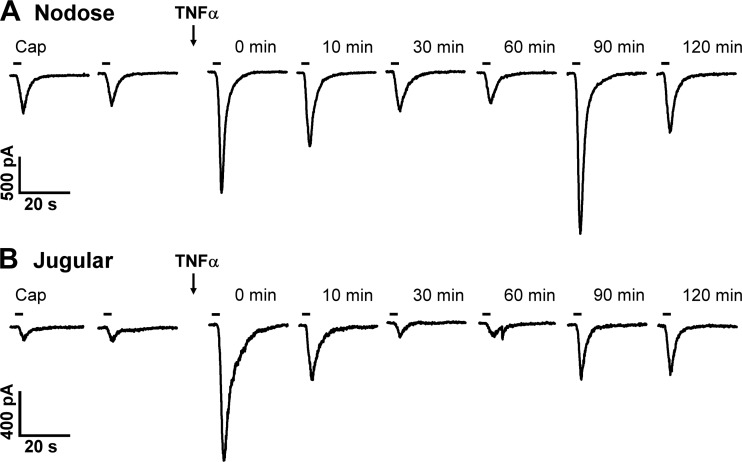
Fig. 2.
Representative experimental records illustrating the inward currents evoked by Cap before and at 0, 10, 30, 60, 90, and 120 min after pretreatment with TNFα in a nodose (A: 18.6 pF) and a jugular (B: 15.1 pF) sensory neuron. Arrows indicate the TNFα (1.44 nM, 9 min) pretreatment. 0 min: the Cap response immediately after the termination of TNFα pretreatment. Cap (0.1 µM, 3 s) application was marked by the horizontal bar; two consecutive Cap challenges before TNFα pretreatment were separated by 10 min.

