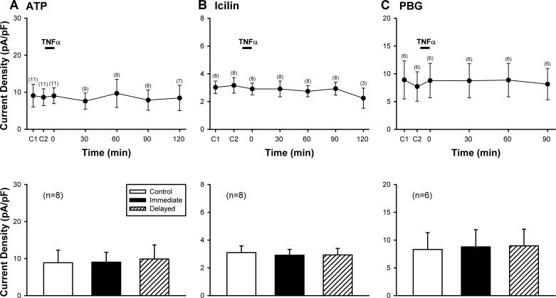
Fig. 5.
A lack of potentiating effect of TNFα pretreatment when Cap was replaced by non-TRPV1 activators of rat vagal pulmonary sensory neurons. Top: histograms of group data illustrating the current densities evoked by ATP (0.3 or 1 µM, 3 s) in A, icilin (100 µM, 4 s) in B, and phenylbiguanide (PBG; 10 µM, 3 s) in C, before and at different time points after TNFα (1.44 nM, 9 min) pretreatment in nodose and jugular sensory neurons, except that ATP responses were only studied in nodose neurons. C1 and C2: control (before TNFα) responses to two consecutive challenges separated by 10 min in A and C; and by 20 min in B. The number of neurons tested at each time point is shown in parenthesis. Bottom: comparisons between responses to the same challenges (ATP, icilin or PBG) between control, immediate (0 min) and delayed (60–90 min) phases after the TNFα pretreatment; these are the same time points when the responses to Cap were potentiated by TNFα as shown in Fig. 3. Data are means ± SE.

